Use the body size and shape as the reference when comparing crystal desk pieces.
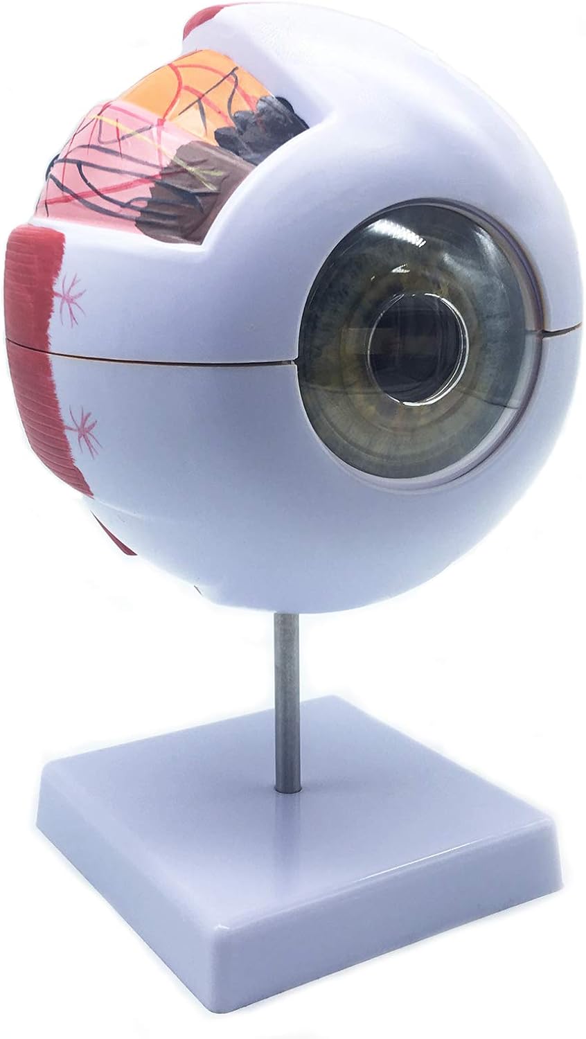
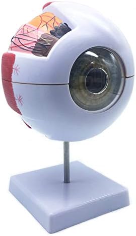
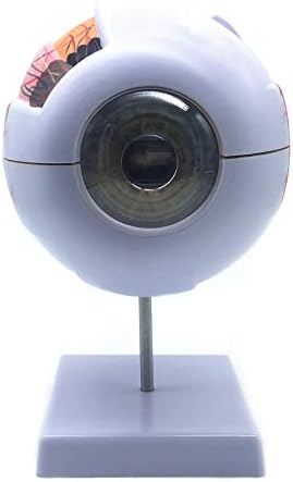
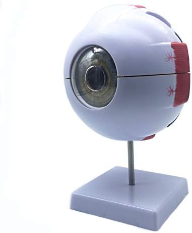

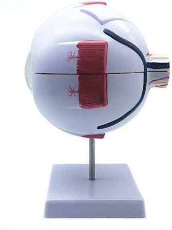
XINDAM 6X Enlarged Human Eye Anatomical Model - Accurate Anatomy for Science Classroom Study, Display, Teaching Medical Model
$29.88
- Material:The model is made of polyvinyl chloride (PVC) plastic, which is corrosion resistant, lightweight, and has high strength.
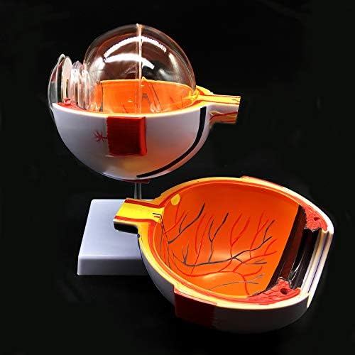
- 6X Enlarged Human Eye Model on base for use in for patient education or anatomical study.You can clearly see all the main anatomical structures of the human eye.The accuracy of this anatomy human eye is the perfect study tool for anatomy students.
- Functional characteristics: the model is used to display the anatomical structure of human eyeball, such as the three layers of outer membrane (outer membrane, media and intima) of eyeball wall and the main refractive bodies, lens and vitreous body filled inside.
- The model includes the labeled diagram for the main parts of the eye.
Crystal guide
Crystal product guide
Use the product images, size notes and base details as the reference point before ordering.
Confirm the artwork area, line count and logo fit before you place a custom order.
Some pieces include a base. Match the base type before you add the product to cart.
Paperweights and ornaments are sized for desks, bookshelves and display cabinets.
Check the spelling, punctuation and logo placement before we finalize the quote.
Clean crystal pieces with a soft cloth and avoid harsh abrasion around the etched area.
Crystal details
CUSTOMER REVIEWS
What customers say
Reviews collected from external sales channels
No review text is available for this product.